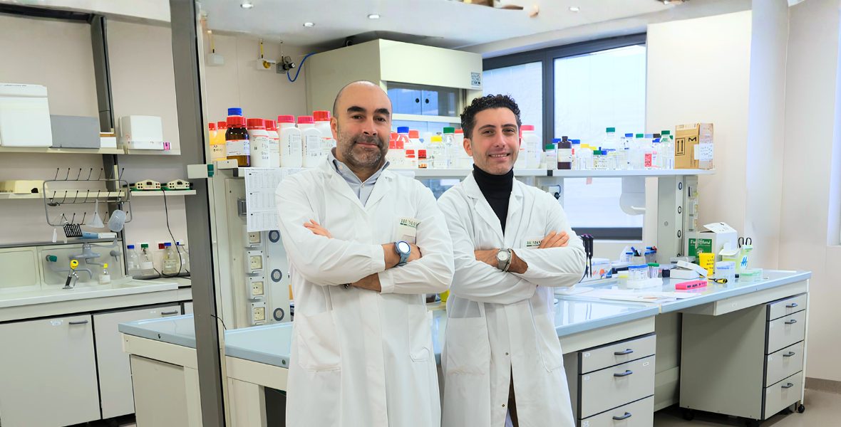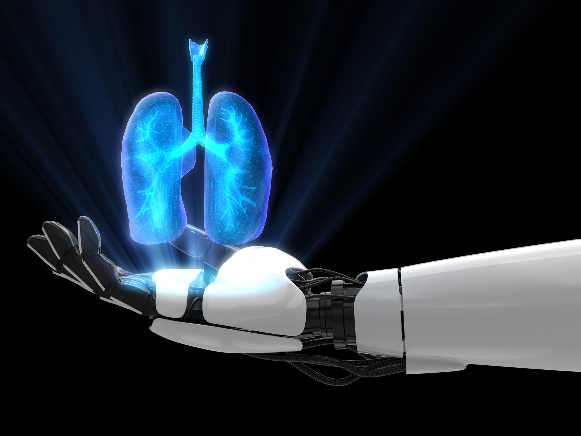
Artificial intelligence can be used to improve lung cancer screening and diagnosis. This is the discovery made by the scientists from Humanitas Department of Diagnostic Imaging and the Department of Biomedical Sciences of Humanitas University, under the direction of Prof. Arturo Chiti, Head of the Nuclear Medicine Operational Unit.
Can artificial intelligence help finding lung cancer and understanding its nature?
Artificial intelligence systems can be used to process imaging results and cross-reference them with other data. The question underlying our studies is whether there is a correlation between tumor genetics and CT and PET images, which allows us to understand if the tumor is more or less aggressive and if it shows genetic anomalies or variations. With our first study, we have developed an analysis which, using the algorithms, proves the correlation between some of these variables.
What data are crossed?
We work with CT and PET images by crossing them with data from blood tests. At the moment, diagnostic imaging is normally used to identify alterations, then a biopsy is performed and the tissue is analyzed. The therapy is decided based on these elements. In the future, the process could change: we will be able to use the images and other data, obtained from a blood sample, correlate them through an artificial intelligence algorithm, and set the therapy based on the results. It is what we call an ‘image biopsy’.
How long did the study last and how many images are needed to “train” the algorithm?
The first project lasted three years and ended in December 2019. The second, based on the concept of ‘biopsy for images’ has recently started, we will have the results in the coming years. For a correct analysis, hundreds of images must be submitted to the machine. So far we have tested a model on a limited number of patients, but the results are encouraging.
Is it already possible to use algorithms to identify lung injury in a screening phase?
We have created an artificial intelligence algorithm that “reads” CT images of patients screened for lung cancer. This is another project that we have completed, we hope to introduce the tool in clinical practice in the near future.
How does it work?
The machine reads the images and detects the possible nodules. It is trained to be very sensitive and to report anything that could constitute a suspicious alteration. The radiologist will then examine the selected images and determine what the alteration is.
What does the new ‘biopsy imaging’ project involve?
The aim of the project is to integrate data from CT and PET images, from circulating DNA and from medical records, to identify the best therapy for lung cancer patients. The integration of the data will be done with artificial intelligence algorithms developed by Humanitas, which will allow to divide the patients into groups, according to the characteristics of the disease. This will lead to choose the most effective therapies.
Starting from the results of the first study, we started a second project that aims to research prognostic factors on lung cancer. The ambition is to develop a software that not only indicates if there are injuries, but can also provide information about their nature. By crossing the data on the nature of the lung lesions seen in the images, with data obtained from a blood sample, in which we search for the DNA that the tumor releases, the algorithm allows us to place patients in a risk class and customize the therapy.