Our Products
Advanced 3D printing and biofabrication offer a complete range of medical solutions, from patient-specific anatomical models and custom surgical guides that enhance planning and procedural accuracy, to high-fidelity phantoms that replicate tissue structures for hands-on training and preclinical research. Biofabricated scaffolds and hydrogel constructs further extend these capabilities by mimicking the properties of human tissues, supporting applications in regenerative medicine, drug delivery and disease modeling, all combining precision, realism, and practical impact across healthcare and life sciences.


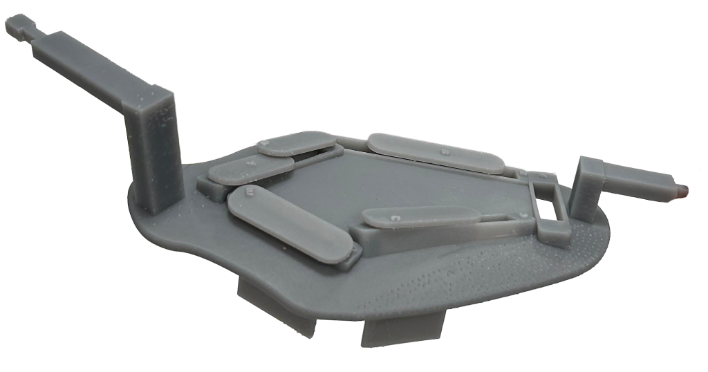
Anesthetic Drug Safety Tray
A 3D-printed solution designed to reduce medication errors through intuitive spatial organisation and ISO-compliant coding.
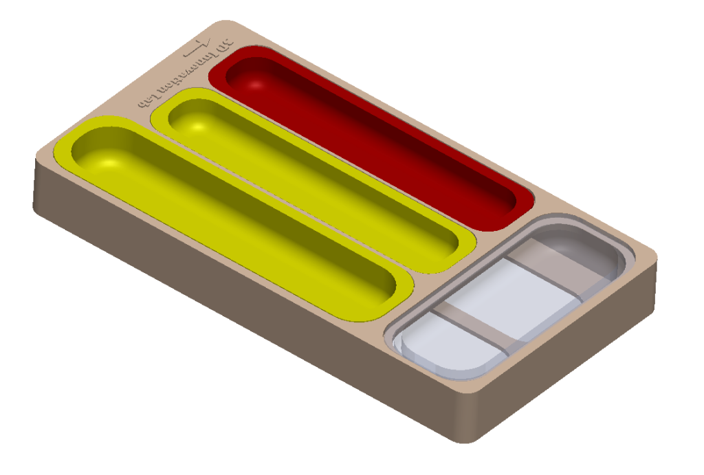
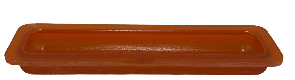
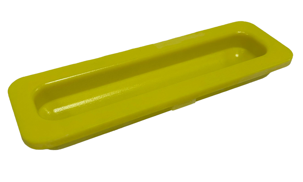
Medication errors in anesthesia are a rare but significant cause of adverse events, often exacerbated by high cognitive demands and challenging environmental conditions such as low lighting or emergencies. The 3D-printed anesthesia tray addresses these challenges by providing a physical tool to standardize the layout of the anesthesia cart. Developed by the 3D innovation lab in partnership with clinical anesthesiologists, this tray features designated, color-coded slots for opioids, neuromuscular blockers, and sedatives, promoting intuitive use and workflow efficiency. Unlike generic commercial options, this model is designed specifically for clinical needs, ensuring high functionality at a sustainable cost.
Key Benefits
- Error reduction: mitigates the risk of drug administration errors by providing clear, fixed locations for specific drug classes
- Enhanced efficiency: streamlines the workspace, allowing anesthesiologists to focus on patient care rather than medication organization
- Visual clarity: utilizes high-contrast color coding to ensure rapid identification of agents
Features
Safety
Reduces human error via visual aids and standardized disposition.
Standards
Color-coding follows ISO 26825:2008 and JCI labeling guidelines.
Design
Compartmentalized layout optimized for routine intraoperative agents.
Production
Sustainable, cost-effective 3D printing technology.
Applications
- Routine anesthetic administration
- Operating room safety & organization
- Cognitive load reduction in critical care
Technical Specifications
Breast Phantom (Resin Based)
High-fidelity patient-specific model for advanced oncoplastic surgery and microsurgical reconstruction.
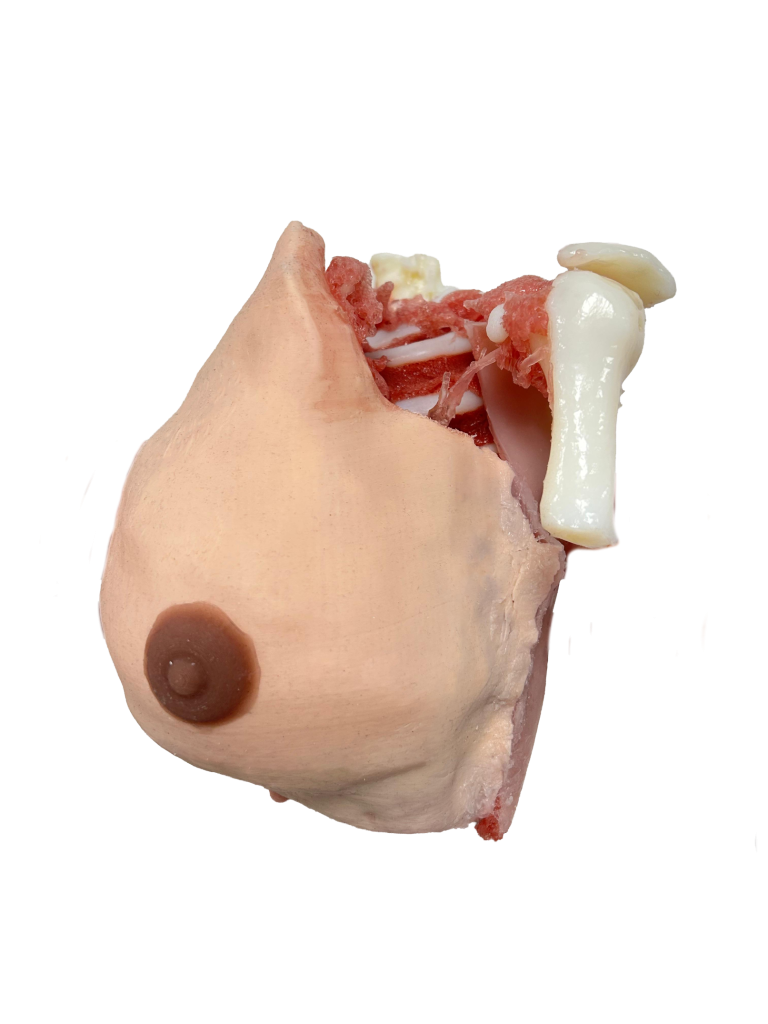
The breast pathological phantom is a high-fidelity surgical simulator designed to overcome the limitations of standard synthetic models and cadaver labs. Specifically engineered for the “complex” configuration, it integrates a rigid skeletal structure with realistic soft tissues, bridging the gap between theoretical knowledge and operating room proficiency. Its patient-specific design allows surgeons to practice on realistic pathologies, offering a valid platform for both skill acquisition and advanced case planning.
Key Benefits
- Deep anatomy & skeletal structure: unlike simple models, this complex unit features a rigid ribcage, sternum, and complete muscular layers (pectoralis major and intercostals), allowing for realistic deep tissue manipulation and feedback.
- Vascular precision: includes an intricate vascular network down to 3 mm in diameter, specifically validated for training in perforator identification and dissection (e.g., aicap).
- Hybrid haptic fidelity: the combination of 3d printed multi-materials and silicone post-processing ensures distinct tactile feedback between glandular tissue, adipose tissue, and tumors, mimicking the resistance of live surgery.
Features
Anatomy
Rigid ribcage, sternum, pectoralis major & intercostal muscles, glandular tissue, adipose tissue, and vascular system.
Materials
Multi-material 3d printing for internal differentiation combined with soft silicone casting for realistic skin and muscle texture.
Customisation
Patient-specific pathologies and anatomies generated directly from high-resolution mri segmentation.
Intended Use
Educational and medical simulation.
Applications
- Tumor excision
- Wide local excision
- Mastectomy flaps simulation
- Perforator flap dissection (aicap)
- Pre-operative surgical planning
- Microsurgical simulation
Technical Specifications
Breast Phantom (Silicone Based)
High-fidelity pathological breast model for surgical simulation.
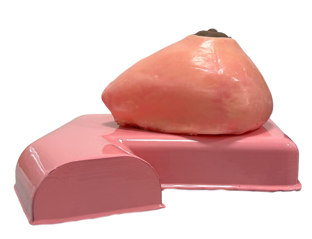
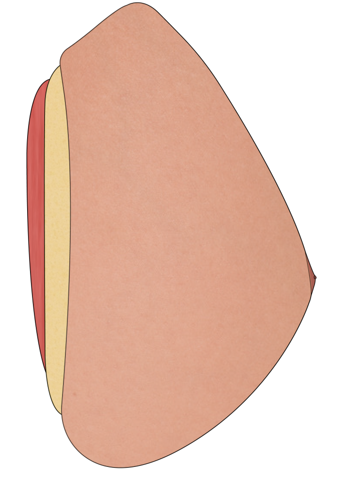
This pathological breast anatomical model is designed to provide a high-fidelity surgical training experience. Manufactured entirely from a variety of silicones, specifically selected to best suit the requirements of each layer, the phantom accurately replicates the resistance, elasticity, and tactile feedback of real biological tissues.
The breast phantom is a comprehensive training tool designed for every surgical operation, including tumor resection, deepithelialization, and mastopexy procedures. The model features a complex anatomical composition that includes the epidermis, dermis, adipose tissue, thoracic muscle, and nipple.
Key Benefits
- High-fidelity anatomical replication & layer separation: the model accurately replicates the anatomy of the female breast. It includes representations of all key anatomical layers, including a specific epidermis (0.40mm), dermis (0.46mm), adipose tissue, thoracic muscle, and nipple. The precise calibration of these layers allows trainees to practice advanced deepithelialization techniques, essential for mastopexy and reconstructive flaps, by realistically separating the epidermal layer from the underlying dermis.
- Pathological simulation & tumor resection: the model showcases a realistic pathological scenario by incorporating a solid tumor made of 3d-printed pla. It helps surgical students and professionals understand the tactile feedback of masses within the tissue, allowing for effective practice of tumor identification and resection operations.
- Realistic tissue behavior: the model displays the resistance and elasticity typical of biological tissues. This feature is crucial for mastering surgical skills such as incision, layer-by-layer dissection, and suturing, ensuring that the trainee experiences the correct haptic feedback required for safe and effective surgery.
Features
Material Composition
Multi-layer silicone structure for realistic tactile feedback.
Skin Layers
Includes epidermis (0.40mm) and dermis (0.46mm).
Internal Anatomy
Adipose tissue, thoracic muscle, nipple.
Intended Use
Educational and medical simulation.
Applications
- De-epithelialization
- Tumor resection
- Mastopexy
- Incision techniques
- Suturing
Technical Specifications
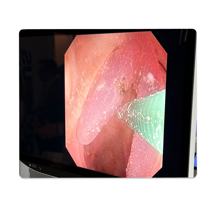
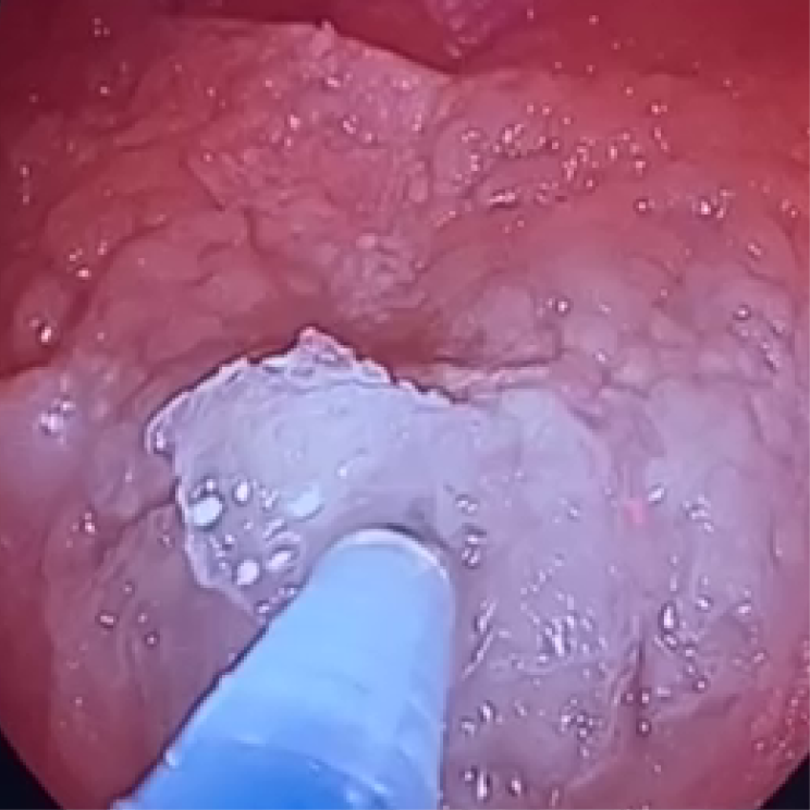
Colon Phantom
High-fidelity, pluri-pathological & injectable model for endoscopic training and AI validation.
The Colon Phantom is a pluri-pathological anatomical model designed for realistic endoscopic simulation. It features a unique 3-layer replica (Inner, Mucosa/Submucosa, Muscle) that overcomes the limitations of single-material models.
Key Benefits
- Multi-use durability: engineered for longevity, offering an extended shelf life compared to biological or gel-based alternatives
- Injectable & realistic: specifically designed for clinical training, it allows for realistic bleb formation during injection procedures
- Pluri-pathological for AI: integrated with different types of lesions, making it an ideal tool for training and validating AI diagnostic algorithms
Features
Anatomical Structure
3-layer replica: inner surface, mucosa/submucosa, muscle.
Pathology Profile
Pluri-pathological: features different types of lesions.
Durability
Multi-use with extended shelf life.
Intended Use
Educational and medical simulation.
Applications
- Clinical procedural training (endoscopy)
- Submucosal fluid injection (bleb formation)
- AI training & validation
- Lesion detection & diagnosis
Technical Specifications
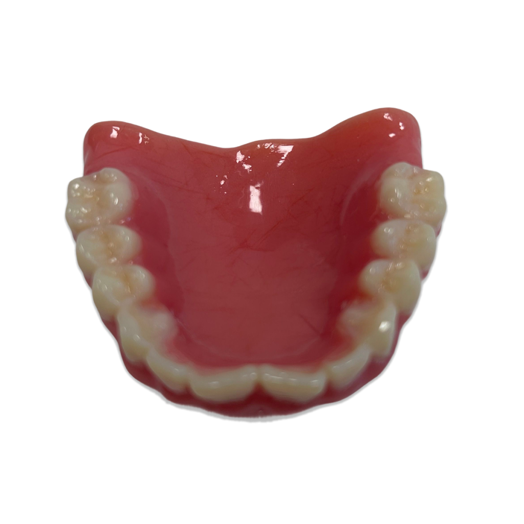
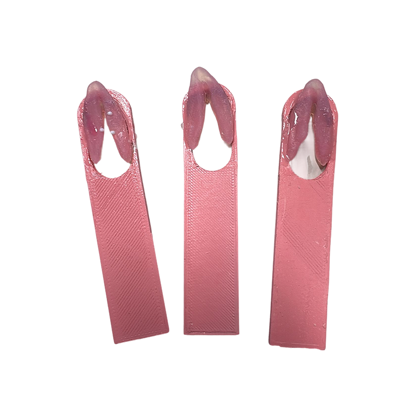
Dental Training Phantom
High-fidelity model specifically designed for dental surgery, implant placement and tooth extraction training.
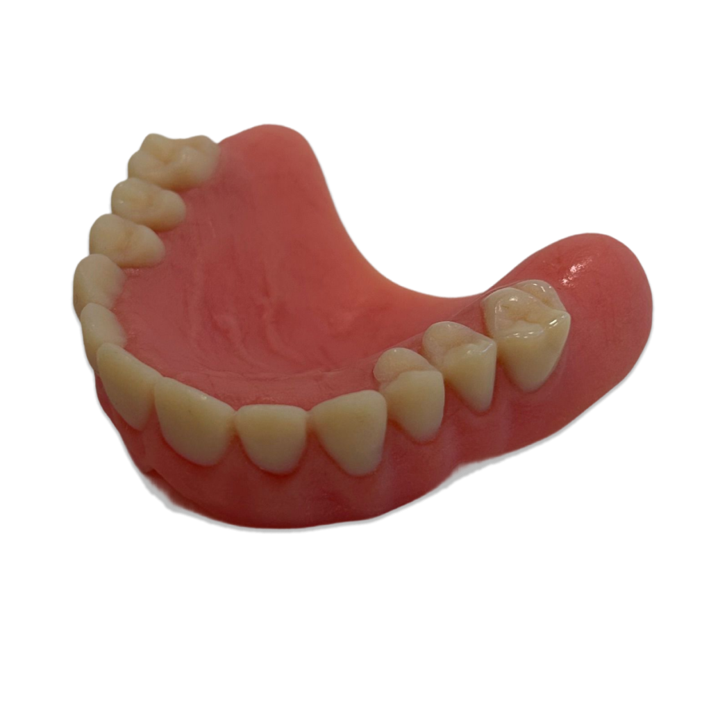
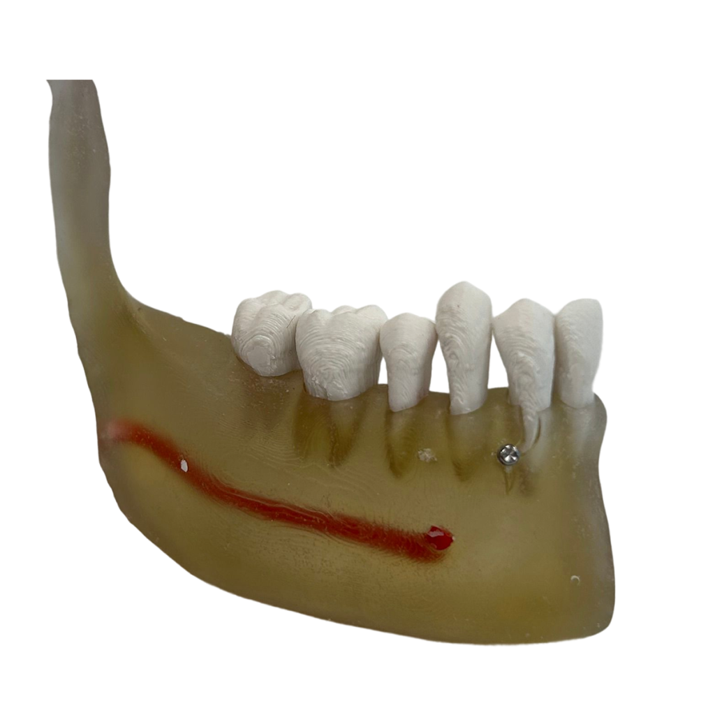
The Dental Training Phantom is a validated high-fidelity simulator that addresses the demand for highly realistic surgical simulation. Unlike standard rigid models, it utilizes advanced multi-material PolyJet 3D printing technology to replicate the complex internal and external anatomy of the human jaw, allowing for the realistic execution of complex dental interventions.
Key Benefits
- Internal Anatomy Visibility: Engineered with simulated translucent structures, such as the mandibular nerve canal, allowing for precise “safe-zone” surgical control
- True-to-Life Haptics: The gingival material is specifically formulated to match the exact resistance, flexibility, and behavior of human soft tissue, essential for incision and suturing
- Anatomical Accuracy: Replicates realistic root structures, alveolar bone morphology, and a fully extractable dentition to provide a true-to-life training experience
Features
Anatomical Structure
Specialized blend of resins with varying consistencies and colours replicating bone, teeth and gums.
Structure & Logic
Layered Architecture: ensures correct interaction for tooth extraction and implant drilling.
Haptic Feedback
Realistic Gingiva: engineered to match human resistance to scalpel incisions and suturing.
Intended Use
Educational and medical simulation.
Applications
- Tooth Extraction Training
- Dental Implant Positioning
- Mandibular Nerve Management
- Soft Tissue Incision and Suturing
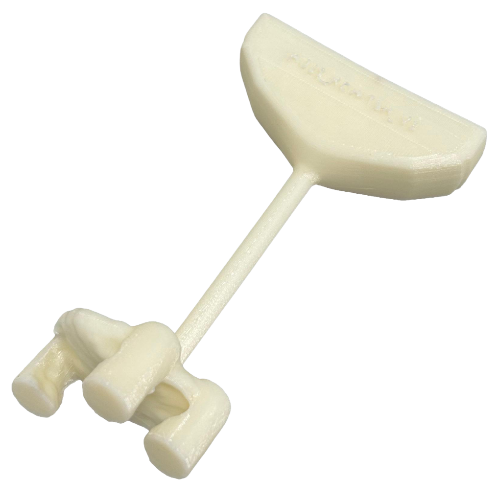
Optional Accessory
Phantom Head/Articulator Support: designed to hold the dental phantom in place and enhance the fidelity of the surgical setup.
Technical Specifications
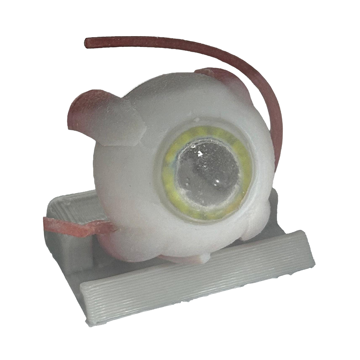
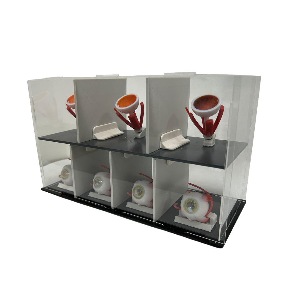
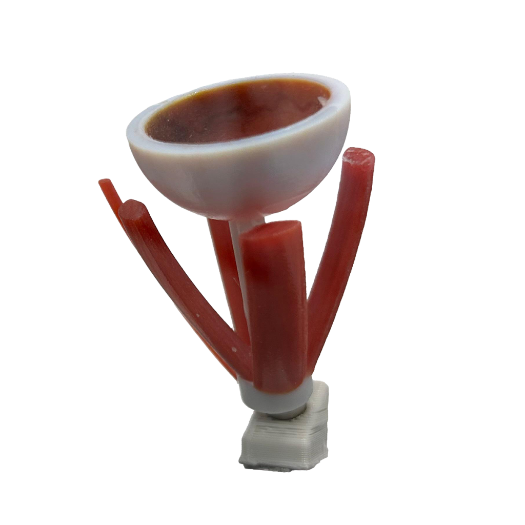
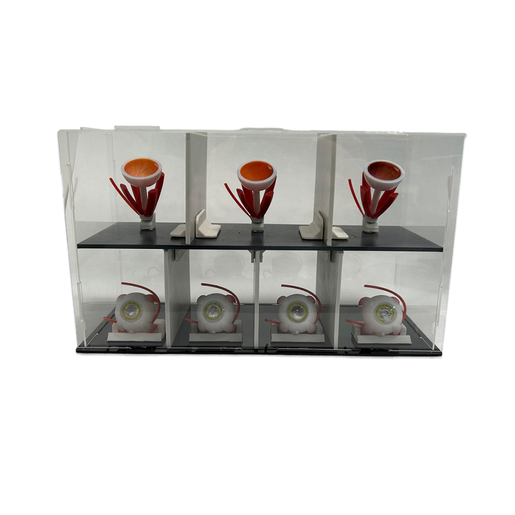
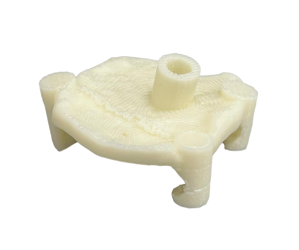
Eye Phantom
High-fidelity model specifically designed for retinal surgery and subretinal injection training.
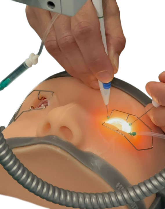
The 3D-eye phantom is a validated high-fidelity simulator that fills the gap in posterior segment training. Unlike standard anterior-only models, it allows for the realistic execution of complex subretinal injections with accurate fluid dynamics.
Optional accessory: facial mask support, designed to hold the eye phantom in place and enhance the fidelity of the surgical setup.
Patent No. 102025000000543
Key Benefits
- Realistic bleb formation: the unique layered structure allows for tissue lifting, accurately mimicking the behavior of the retina during fluid injection.
- True-to-life haptics: the sclera material is mechanically tested to replicate the exact resistance to puncturing of the human eye, essential for trocar insertion.
- Anatomical accuracy: includes critical references (capillaries, optic nerve, fovea) and an intraoperative lens for optimal visualization during surgery.
Features
Anatomical Structure
Specialized blend of resins with varying consistencies and colours.
Structure & Logic
Layered architecture: ensures correct interaction for bleb formation.
Haptic Feedback
Realistic sclera: engineered to match human resistance to puncturing.
Intended Use
Educational and medical simulation.
Applications
- Subretinal fluid injection (bleb formation)
- Epiretinal peeling
- Trocar insertion training
- Posterior segment surgery simulation
Technical Specifications
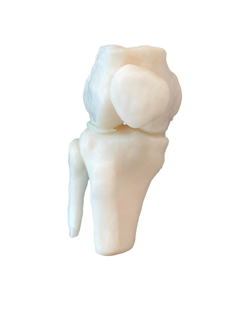
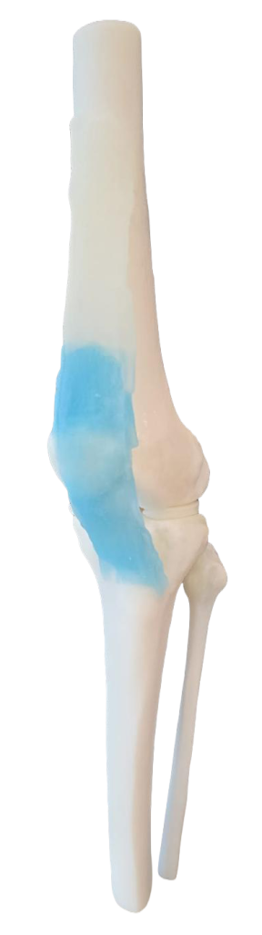
Knee Phantom
A hybrid simulator combining rigid skeletal accuracy, flexible ligaments and a zippered soft-tissue envelope for versatile orthopedic training.
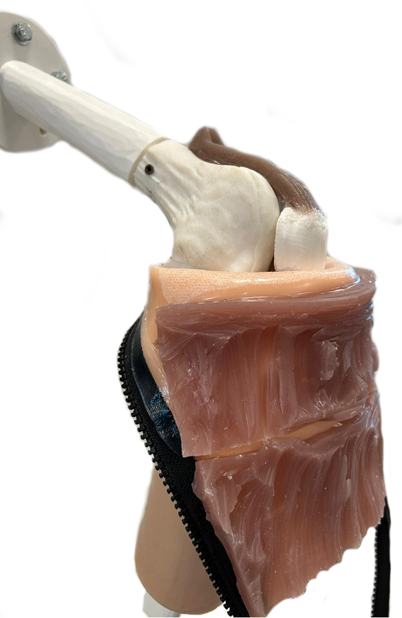
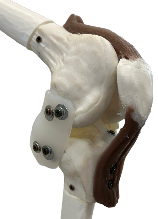
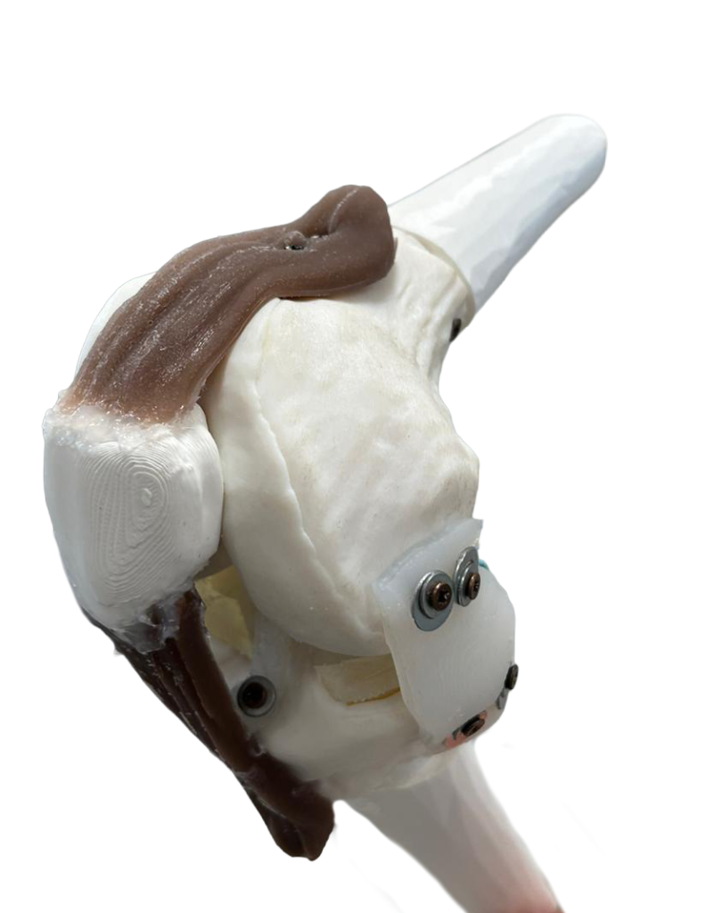
The High-Fidelity Knee Phantom bridges the gap between virtual simulation and clinical practice. Specifically designed for sports medicine and orthopedic education, this model offers a risk-free environment for skill development. The phantom features a sophisticated hybrid construction: rigid 3D-printed bones provide anatomical landmarks, while flexible ligaments and a realistic meniscus allow for authentic physiological feedback. A key feature is the removable soft-tissue envelope with a zipper, which facilitates the explanation of knee structures and allows trainees to visualize the correlation between external landmarks and internal anatomy. Securely mounted on a stand, it is the ideal tool for practicing arthroscopic triangulation, injection techniques, and general joint manipulation.
Features
Anatomy
Accurate bone anatomy (femur, tibia, patella) with functional ligaments and menisci.
Design
Removable soft-tissue sleeve with zipper for easy visualisation of internal structures.
Stability
Stand-mounted design ensures stability during complex maneuvers.
Simulation
Suitable for arthroscopy, injections, and physical manipulation without patient risk.
Applications
- Arthroscopic Meniscal Repair
- Intra-articular injections
- Orthopedic Joint Manipulation
Technical Specifications
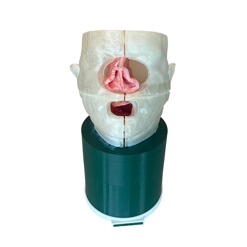
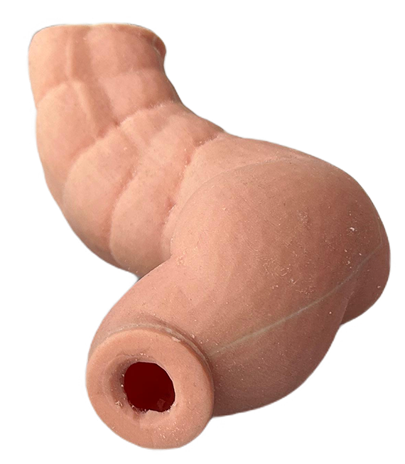
Larynx & Airway Pathological Phantom
High-fidelity patient-specific model for advanced endolaryngeal surgery and airway management training.
The Larynx & Airway Pathological Phantom is a high-fidelity surgical simulator designed to overcome the limitations of standard synthetic models and cadaver labs. Specifically engineered for endolaryngeal and laser-based procedures, it integrates a realistic respiratory tract with pathological tissue, bridging the gap between theoretical knowledge and operating room proficiency. Its patient-specific design allows surgeons to practice on realistic pathologies, offering a valid platform for both skill acquisition and advanced case planning.
- Hybrid Haptic Fidelity: The combination of 3D printed multi-materials and specialized post-processing ensures distinct tactile feedback between mucosal surfaces, vocal folds, and tumors, mimicking the resistance of live surgery. Includes replaceable vocal cord modules (consumables) for repetitive surgical practice
- Deep Anatomy & Airway Structure: Unlike simple models, this complex unit features a complete path from the nose to the trachea, including cartilaginous rings and laryngeal musculature, allowing for realistic instrument navigation and feedback
- Laser-Tissue Precision: The model utilizes proprietary materials validated for laser surgery, responding to energy precisely like real mucosa and tumors, supporting the practice of delicate perforator-level identification and resection
Features
Anatomical Structure
Nose, pharynx, larynx with pathological vocal cords, trachea with cartilaginous rings and laryngeal muscles.
Materials
Multi-material 3D printing for structural cartilages combined with laser-responsive soft polymers for physiological tissue reproduction.
Customisation and Intender Use
Patient-specific pathologies and vocal cord lesions generated from high-resolution clinical imaging. Educational and medical simulation.
Applications
- Vocal cord tumor excision
- Endoscopic microsurgery training
- Laser tissue interaction practice
- Airway management and intubation
Technical Specifications
Pathological Eye
A visual tool designed to enhance patient understanding of eye conditions for clear and effective communication.

The Pathologic Eye Model is a highly realistic visual tool developed by the Humanitas University 3D Innovation Lab to bridge the gap in patient communication.
Unlike mechanical simulators, this model prioritizes high visual fidelity to help clinicians effectively explain diagnoses, treatments, and the progression of various ocular diseases.
This model is intended for educational and communication purposes only and does not replicate the mechanical properties of human tissue for surgical training.
Key Benefits
- High Visual Realism: features a true-to-life appearance that accurately replicates the full anatomical structure of the human eye for visual simulation.
- Comprehensive Anatomical Composition: includes all key structures such as the extraocular muscles, optic nerve, sclera, cornea, and retina.
- Multiple Pathology Representation: designed to support the explanation of a wide range of conditions, specifically focusing on corneal, retinal, and muscular.
Features
Anatomical Structure
Complete reproduction of the eye, including the retina, cornea, sclera, optic nerve, and extraocular muscles.
Visual Fidelity
High realism and true-to-life appearance designed for clear visual simulation of eye conditions.
Pathology Support
Specialized representation of retinal, corneal, and muscular conditions to support clinical explanations.
Intended Use
Dedicated tool for patient educationand medical communication.
Applications
- Patient education in ophthalmology clinics to enhance understanding of eye conditions
- Medical consultations to clearly explain diagnoses and treatment pathways
- Clinical communication support for illustrating disease progression
- Visual simulation of complex ocular pathologies for effective patient-doctor dialogue
Technical Specifications
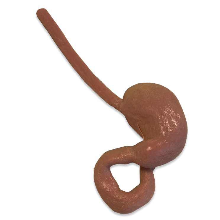
Pathological Heart
A multimaterial, soft-tissue heart model designed to simulate structural pathologies with lifelike haptic feedback.
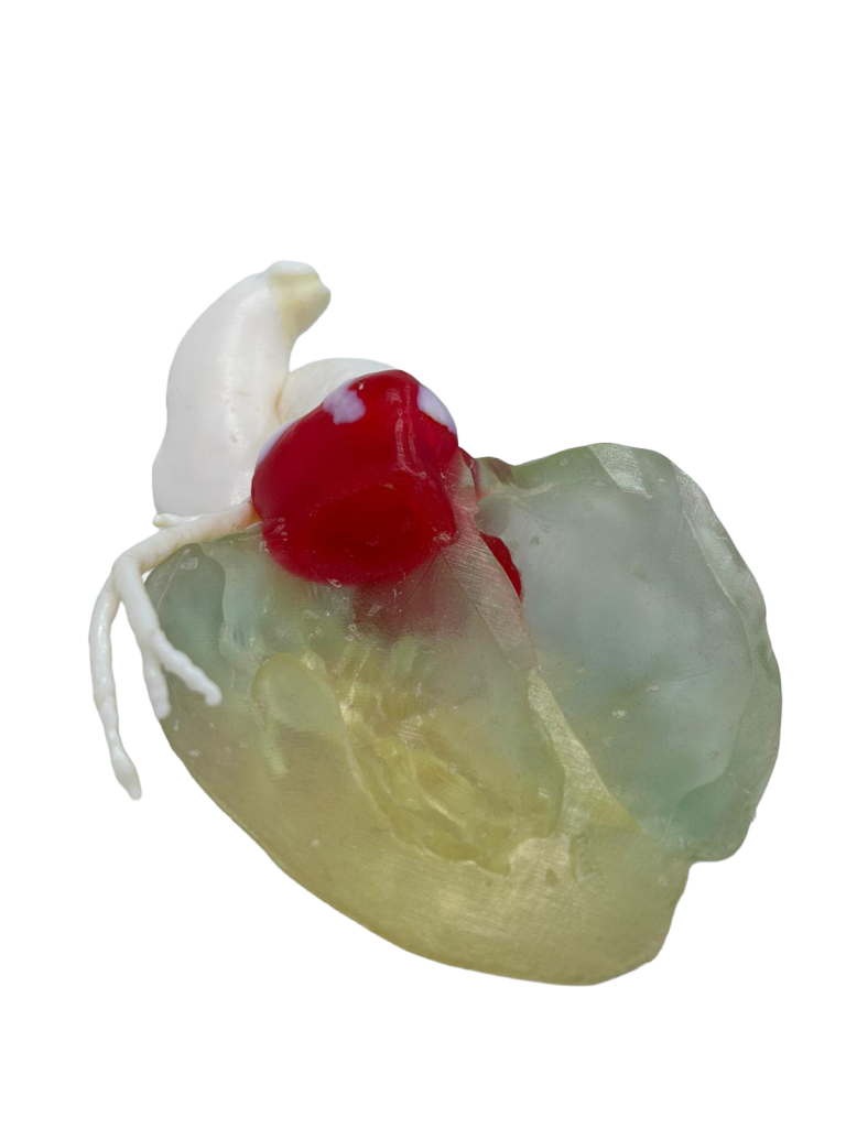
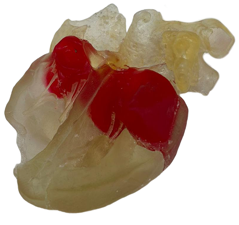
The 3D-printed pathological heart represents a leap forward in cardiac simulation.
The combination of clear, flexible resins for the heart walls and distinct colors for pathologies (such as tumors or septal defects) allows for unparalleled understanding of complex geometries. It is the ideal tool for surgeons practicing intricate procedures and for researchers testing the fit and deployment of new cardiac devices.
Features
Anatomy
Complete cardiac structure including atria, ventricles, major vessels, and specific pathological defects.
Materials
A blend of soft and rigid resins to replicate tissue compliance.
Visualisation
Translucent walls allow full visibility of internal pathologies and device interaction.
Pathology
Includes common structural anomalies such as septal defects or intracardiac masses.
Applications
- Structural heart disease interventions
- Pre-operative surgical planning
- Hemodynamic simulation
Technical Specifications
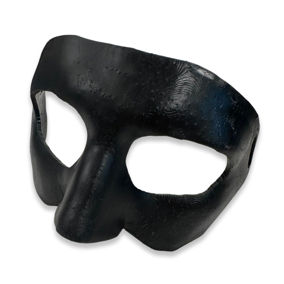
Patient-Specific Nasal Orthosis
Custom nasal orthoses engineered from 3D facial scan to support your recovery and safe return to sports.
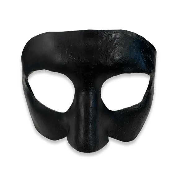
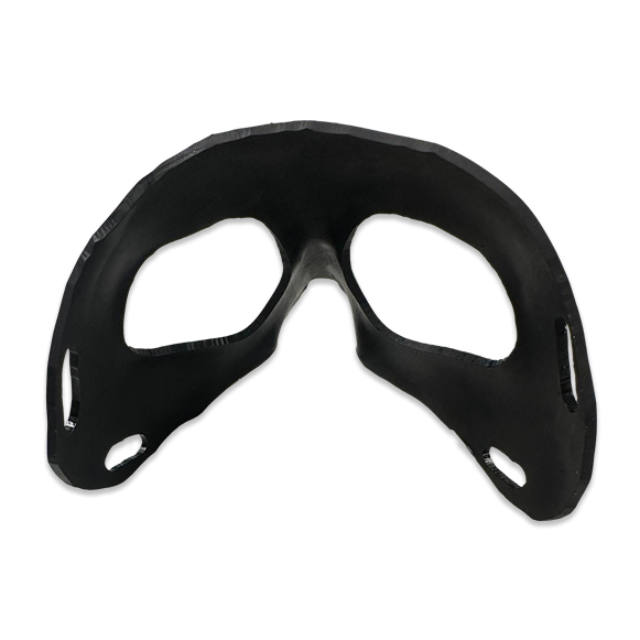
Our Patient-Specific Nasal Orthoses redefine the standards of protection and comfort in facial trauma recovery. By translating a high-resolution 3D facial scan into a physical, ergonomic barrier, these custom-made masks eliminate the limitations of generic, off-the-shelf devices. Specifically engineered for the management of nasal fractures and post-operative care, our orthoses feature a unique structural design that protects the nasal pyramid by safely distributing impact energy to the surrounding forehead and cheekbones. This is critical for both conservative rehabilitation and a safe return to high-impact sports.
Manufactured from lightweight, biocompatible 3D-printed resin, the perfect anatomical fit ensures maximum stability and an entirely unobstructed field of vision, ultimately leading to improved patient compliance and a safer recovery.
Features
Design
Generated from 3D facial scans for a perfect anatomical fit.
Function
Protects the nasal pyramid by safely distributing impact energy.
Material
Biocompatible,lightweight 3D-printed resin safe for prolonged skin contact.
Optimized Stability
Custom-contoured openings ensure a completely unobstructed field of vision.
Applications
- Nasal Fracture Management & Recovery
- Post-Operative Protection (Rhinoplasty)
- Sports Traumatology
- Maxillofacial Trauma Management
Technical Specifications
Patient-Specific Surgical Guides
Custom 3D-printed guides featuring high-precision slots and cylinders for exact drilling, cutting and implant positioning.
Our Patient-Specific Surgical Guides (PSI) redefine intraoperative precision. By translating virtual pre-operative planning into physical reality, these tools eliminate guesswork in the operating room. Specifically for Hard Tissue applications, our guides are equipped with integrated cylinders and slots that dictate the exact angle and depth for saws and drills. This is critical for procedures such as complex shoulder arthroplasty or correcting glenoid deformities. Whether for precise bone resection or soft tissue management, the “snap-on” fit reduces surgical time and reliance on intraoperative fluoroscopy, ultimately leading to better patient outcomes and mechanical stability.
Medical device class IIA compliance to the MDR 2017/745
Features
Design
Generated from patient CT imaging for a perfect anatomical fit.
Function
Accurate drilling trajectories and cutting planes.
Material
Biocompatible, sterilizable materials compliant with ISO 13485:2016.
Optimized Stability
Ensures accurate plan translation and optimal implant alignment.
Applications
- Bone arthroplasty & deformity
- Bone tumor resection & reconstruction
- Management of bone loss & malunions
- Soft tissue oncological margins
Technical Specifications
Stomach Phantom
High-Fidelity, Pluri-Pathological & Injectable Model for Endoscopic Training and AI Validation.
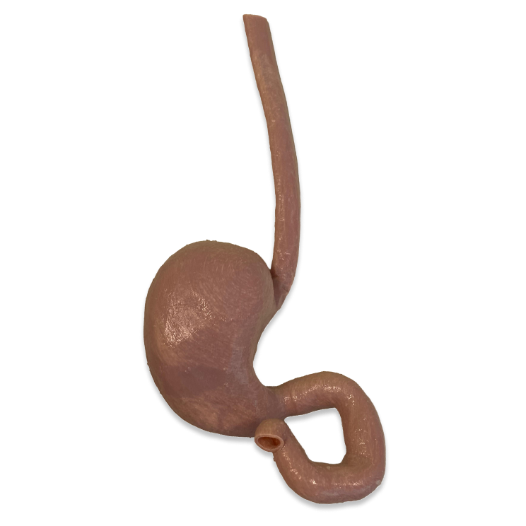
The Stomach Phantom is a pluri-pathological anatomical model designed for realistic endoscopic simulation. It features a unique 3-layer replica (Inner, Mucosa/Submucosa, Muscle) that overcomes the limitations of single-material models.
Key Benefits
- Injectable & Realistic: specifically designed for clinical training, it allows for realistic bleb formation during injection procedures
- Pluri-Pathological for AI: integrated with different types of lesions, making it an ideal tool for training and validating AI diagnostic algorithms
- Multi-Use Durability: engineered for longevity, offering an extended shelf life compared to biological or gel-based alternatives
Features
Anatomical Structure
3-Layer Replica: inner surface, mucosa/submucosa, muscle.
Pathology Profile
Pluri-pathological: features different types of lesions.
Durability
Multi-use with extended shelf life.
Intended Use
Educational and medical simulation.
Applications
- Clinical Procedural Training (Endoscopy)
- Submucosal Fluid Injection (Bleb Formation)
- AI Training & Validation
- Lesion Detection & Diagnosis
Technical Specifications
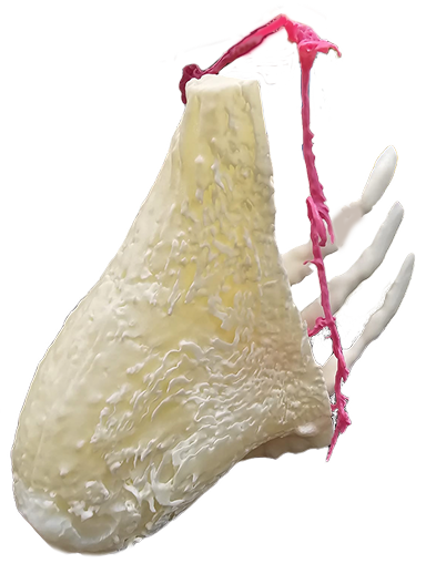
3D-Printed Anatomical Bones
Custom-printed bone models available in rigid FDM for education or high-fidelity PolyJet for surgical simulation and soft tissue integration.
Our 3D-Printed Anatomical Bones category offers tailored solutions for every stage of medical training. Whether you need a durable, anatomically correct femur for a university lecture or a complex spinal column with soft intervertebral discs for surgical rehearsal, we have the technology to deliver. We leverage FDM printing for affordable, rigid models perfect for understanding complex 3D geometries. for advanced simulation, our PolyJet technology creates multi-material phantoms that mimic the biomechanics of real bone and cartilage. These advanced models allow surgeons to practice drilling, reaming, and cutting with lifelike resistance, making them invaluable for pre-operative planning and medical device testing.
Features
Customisation
Patient-Specific bones, printed on request from medical imaging.
Technologies
FDM for durable educational models; PolyJet for realistic surgical simulation.
Soft Tissue
Integrated cartilaginous structures (menisci, discs), ligaments and tendons (PolyJet Only).
Simulation
Realistic response to drilling, sawing and milling for orthopedic training.
Applications
- Anatomy education & training
- Pre-operative surgical planning
- Orthopedic instrumentation testing
- Soft tissue oncological margins