A clinical study conducted at Humanitas demonstrates AI’s ability to distinguish between tumor progression and treatment-induced tissue changes after radiotherapy, paving the way for more precise and less invasive therapeutic decisions.
After radiotherapy treatment for brain metastases, distinguishing between radiotherapy-induced tissue changes and tumor progression remains a critical diagnostic challenge. Conventional diagnostic techniques, such as magnetic resonance imaging (MRI), often fail to provide definitive answers, as the radiological characteristics of the two tissue types are strikingly similar. According to the results of a new study published in Neuro Oncology, Artificial Intelligence (AI) could overcome these limitations: the findings demonstrate AI’s superior ability to differentiate between radionecrosis and tumor progression in patients with brain metastases treated with stereotactic radiosurgery, a highly precise radiotherapy technique.
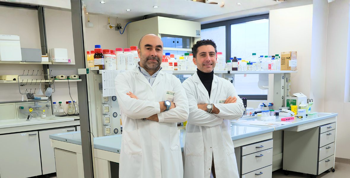
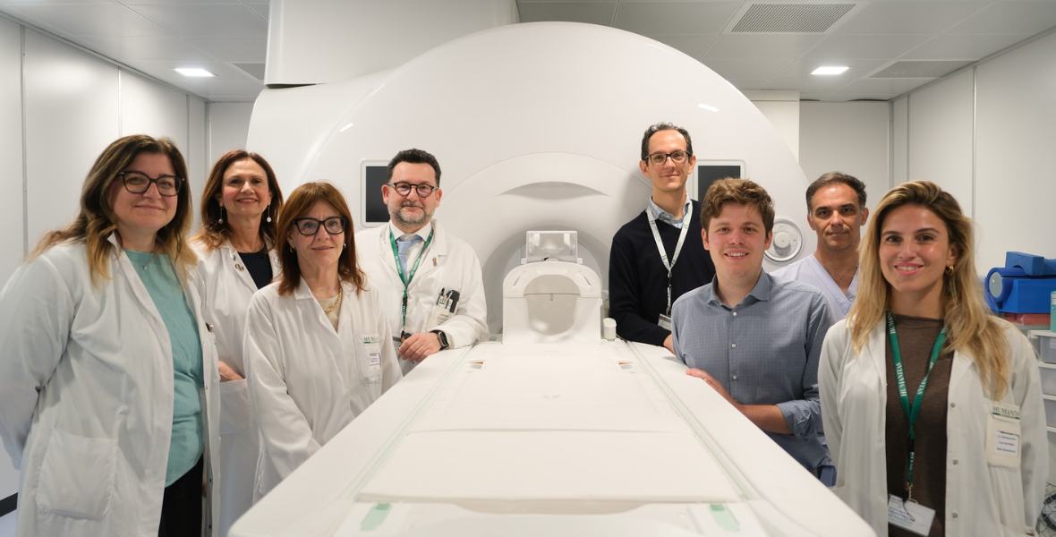
The research, led by a multidisciplinary team of neuroradiologists, oncologists, radiotherapists, and pathologists in collaboration with Humanitas Research Hospital, Università La Sapienza di Roma and University Hospital of Tübingen, was coordinated by Prof. Letterio Politi − Head of Neuroradiology at Humanitas Research Hospital − and Prof. Marta Scorsetti − Head of Radiotherapy and Radiosurgery, both faculty members at Humanitas University.
The researchers retrospectively analyzed 124 brain lesions in patients who had undergone stereotactic radiotherapy and had histological confirmation through biopsy or surgical resection. The study utilized radiomics approaches, which extract quantitative information from medical images, and deep learning models, AI algorithms that leverage artificial neural networks for learning.
The results revealed that Artificial Intelligence algorithms can accurately differentiate radionecrosis from tumor progression. After being trained on clinical cases from the hospital, the algorithm was retrospectively tested on an external patient cohort, allowing for the validation of the approach and generalization of the findings.
“Stereotactic radiosurgery is an effective technique for treating brain metastases,” explains Marta Scorsetti. “However, distinguishing between radionecrosis, a side effect of treatment, and tumor progression can be challenging. Our research demonstrates that AI has the potential to provide a more accurate diagnostic tool, potentially reducing the need for exploratory biopsy or surgical procedures.”
“While further research is needed to validate these models in larger patient populations, the results are promising and suggest that AI could become a valuable tool supporting the work of neuroradiologists today and in the future,” says Letterio Politi, who also leads the Master’s Degree Program in Data Analytics and Artificial Intelligence in Health Sciences (DAIHS), created in collaboration between Humanitas University and Bocconi University to combine biomedical and healthcare expertise with AI, data science, and data analytics skills.
Scorsetti and Politi conclude: “This research represents an important step forward in the field of neuro-oncology and provides further evidence of how advanced technologies and multidisciplinary collaboration can combine to enhance our diagnostic and treatment capabilities, moving closer to precision medicine approaches.”